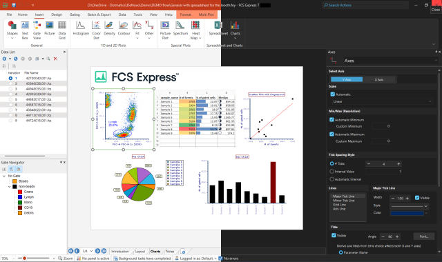
Know how to use Microsoft PowerPoint™? How about Excel™?
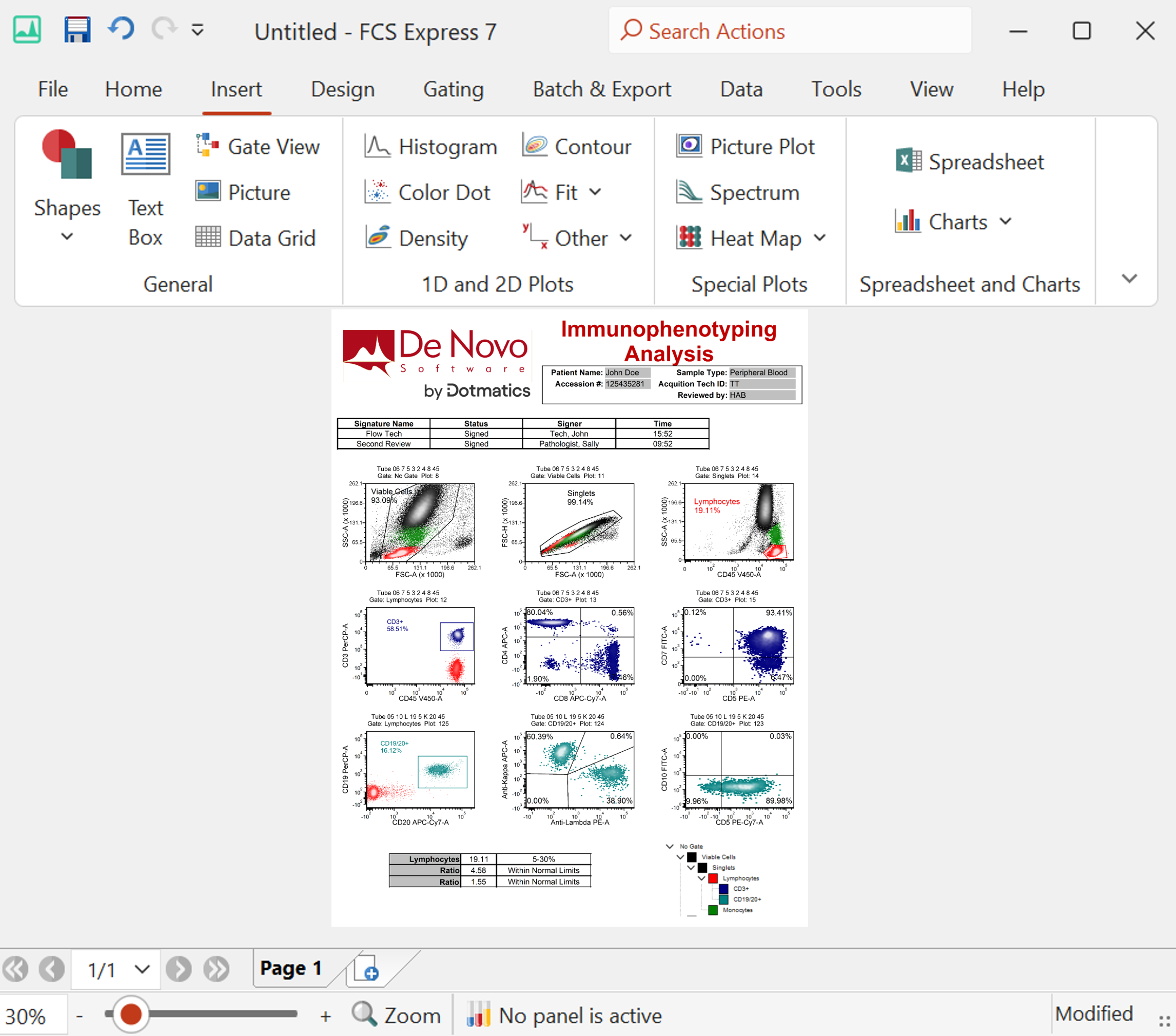
Learning how to use flow cytometry software should not be an impediment to getting results from your data. FCS Express™ is designed to look, feel, and work like many familiar Microsoft Office™ programs so you are already half way to being an expert with the software before you even get started.